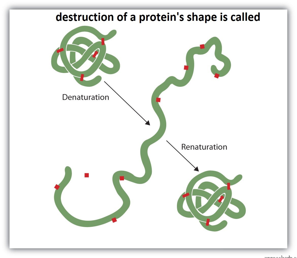
destruction of a protein’s shape is called
Hi dear friends, solsarin in this article is talking about “destruction of a protein’s shape is called”.
we are happy to have you on our website.
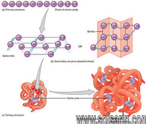
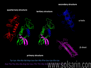
We have seen that each type of protein consists of a precise sequence of amino acids that allows it to fold up into a particular three-dimensional shape, or conformation. But proteins are not rigid lumps of material. They can have precisely engineered moving parts whose mechanical actions are coupled to chemical events. It is this coupling of chemistry and movement that gives proteins the extraordinary capabilities that underlie the dynamic processes in living cells.
In this section, we explain how proteins bind to other selected molecules and how their activity depends on such binding. We show that the ability to bind to other molecules enables proteins to act as catalysts, signal receptors, switches, motors, or tiny pumps. The examples we discuss in this chapter by no means exhaust the vast functional repertoire of proteins. However, the specialized functions of many of the proteins you will encounter elsewhere in this book are based on similar principles.
All Proteins Bind to Other Molecules
The biological properties of a protein molecule depend on its physical interaction with other molecules. Thus, antibodies attach to viruses or bacteria to mark them for destruction, the enzyme hexokinase binds glucose and ATP so as to catalyze a reaction between them, actin molecules bind to each other to assemble into actin filaments, and so on. Indeed, all proteins stick, or bind, to other molecules.
In some cases, this binding is very tight; in others, it is weak and short-lived. But the binding always shows great specificity, in the sense that each protein molecule can usually bind just one or a few molecules out of the many thousands of different types it encounters. The substance that is bound by the protein—no matter whether it is an ion, a small molecule, or a macromolecule— is referred to as a ligand for that protein (from the Latin word ligare, meaning “to bind”).
The ability of a protein to bind selectively and with high affinity to a ligand depends on the formation of a set of weak, noncovalent bonds—hydrogen bonds, ionic bonds, and van der Waals attractions—plus favorable hydrophobic interactions (see Panel 2-3, pp. 114–115). Because each individual bond is weak, an effective binding interaction requires that many weak bonds be formed simultaneously. This is possible only if the surface contours of the ligand molecule fit very closely to the protein, matching it like a hand in a glove (Figure 3-37).
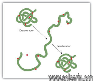
Figure 3-37
The selective binding of a protein to another molecule. Many weak bonds are needed to enable a protein to bind tightly to a second molecule, which is called a ligand for the protein. A ligand must therefore fit precisely into a protein’s binding (more…)
The region of a protein that associates with a ligand, known as the ligand’s binding site, usually consists of a cavity in the protein surface formed by a particular arrangement of amino acids. These amino acids can belong to different portions of the polypeptide chain that are brought together when the protein folds (Figure 3-38). Separate regions of the protein surface generally provide binding sites for different ligands, allowing the protein’s activity to be regulated, as we shall see later. And other parts of the protein can serve as a handle to place the protein in a particular location in the cell—an example is the SH2 domain discussed previously, which is often used to move a protein containing it to sites in the plasma membrane in response to particular signals.
Figure 3-38
The binding site of a protein. (A) The folding of the polypeptide chain typically creates a crevice or cavity on the protein surface. This crevice contains a set of amino acid side chains disposed in such a way that they can make noncovalent bonds only (more…)
Although the atoms buried in the interior of the protein have no direct contact with the ligand, they provide an essential scaffold that gives the surface its contours and chemical properties. Even small changes to the amino acids in the interior of a protein molecule can change its three-dimensional shape enough to destroy a binding site on the surface.
The Details of a Protein’s Conformation Determine Its Chemistry
Proteins have impressive chemical capabilities because the neighboring chemical groups on their surface often interact in ways that enhance the chemical reactivity of amino acid side chains. These interactions fall into two main categories.
First, neighboring parts of the polypeptide chain may interact in a way that restricts the access of water molecules to a ligand binding site. Because water molecules tend to form hydrogen bonds, they can compete with ligands for sites on the protein surface. The tightness of hydrogen bonds (and ionic interactions) between proteins and their ligands is therefore greatly increased if water molecules are excluded.
Initially, it is hard to imagine a mechanism that would exclude a molecule as small as water from a protein surface without affecting the access of the ligand itself. Because of the strong tendency of water molecules to form water–water hydrogen bonds, however, water molecules exist in a large hydrogen-bonded network (see Panel 2-2, pp. 112–113). In effect, a ligand binding site can be kept dry because it is energetically unfavorable for individual water molecules to break away from this network, as they must do to reach into a crevice on a protein’s surface.
Second, the clustering of neighboring polar amino acid side chains can alter their reactivity. If a number of negatively charged side chains are forced together against their mutual repulsion by the way the protein folds, for example, the affinity of the site for a positively charged ion is greatly increased. In addition, when amino acid side chains interact with one another through hydrogen bonds, normally unreactive side groups (such as the –CH2OH on the serine shown in Figure 3-39) can become reactive, enabling them to enter into reactions that make or break selected covalent bonds.
Figure 3-39
An unusually reactive amino acid at the active site of an enzyme. This example is the “catalytic triad” found in chymotrypsin, elastase, and other serine proteases (see Figure 3-14). The aspartic acid side chain (Asp 102) induces the histidine (more…)
The surface of each protein molecule therefore has a unique chemical reactivity that depends not only on which amino acid side chains are exposed, but also on their exact orientation relative to one another. For this reason, even two slightly different conformations of the same protein molecule may differ greatly in their chemistry.
Sequence Comparisons Between Protein Family Members Highlight Crucial Ligand Binding Sites
As we have described previously, many of the domains in proteins can be grouped into families that show clear evidence of their evolution from a common ancestor, and genome sequences reveal large numbers of proteins that contain one or more common domains. The three-dimensional structures of the members of the same domain family are remarkably similar. For example, even when the amino acid sequence identity falls to 25%, the backbone atoms in a domain have been found to follow a common protein fold within 0.2 nanometers (2 Å).
These facts allow a method called “evolutionary tracing” to be used to identify those sites in a protein domain that are the most crucial to the domain’s function. For this purpose, those amino acids that are unchanged, or nearly unchanged, in all of the known protein family members are mapped onto a structural model of the three-dimensional structure of one family member. When this is done, the most invariant positions often form one or more clusters on the protein surface, as illustrated in Figure 3-40A for the SH2 domain described previously (see Panel 3-2, pp. 138–139). These clusters generally correspond to ligand binding sites.
Inhibition of enzymes
Some molecules very similar to the substrate for an enzyme may be bound to the active site but be unable to react. Such molecules cover the active site and thus prevent the binding of the actual substrate to the site. This inhibition of enzyme action is of a competitive nature, because the inhibitor molecule actually competes with the substrate for the active site.
The inhibitor sulfanilamide, for example, is similar enough to a substrate (p-aminobenzoic acid) of an enzyme involved in the metabolism of folic acid that it binds to the enzyme but cannot react. It covers the active site and prevents the binding of p-aminobenzoic acid. This enzyme is essential in certain disease-causing bacteria but is not essential to humans; large amounts of sulfanilamide therefore kill the microorganism but do not harm humans. Inhibitors such as sulfanilamide are called antimetabolites. Sulfanilamide and similar compounds that kill a pathogen without harming its host are widely used in chemotherapy.
Effects of temperature
Enzymes function most efficiently within a physiological temperature range. Since enzymes are protein molecules, they can be destroyed by high temperatures. An example of such destruction, called protein denaturation, is the curdling of milk when it is boiled. Increasing temperature has two effects on an enzyme: first, the velocity of the reaction increases somewhat, because the rate of chemical reactions tends to increase with temperature; and, second, the enzyme is increasingly denatured. Increasing temperature thus increases the metabolic rate only within a limited range. If the temperature becomes too high, enzyme denaturation destroys life. Low temperatures also change the shapes of enzymes.
Enzyme flexibility and allosteric control
The induced-fit theory
The key–lock hypothesis (see above The nature of enzyme-catalyzed reactions) does not fully account for enzymatic action; i.e., certain properties of enzymes cannot be accounted for by the simple relationship between enzyme and substrate proposed by the key–lock hypothesis. A theory called the induced-fit theory retains the key–lock idea of a fit of the substrate at the active site but postulates in addition that the substrate must do more than simply fit into the already preformed shape of an active site.