heterogeneously dense breast tissue
Hello dear friends, solsarin in this article is talking about “heterogeneously dense breast tissue”.
we are happy to have you on our website.
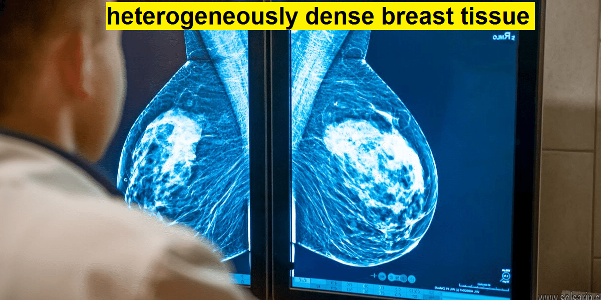
Breast density is a proportional measure of the glandular, connective and fatty tissues within a woman’s breasts. It is most commonly determined using mammography, a diagnostic test that uses low dose x-rays. Having dense breasts is not an abnormal condition; in fact, about half of all women over 40 have dense breasts.
The exact relationship between breast density and breast cancer is still unknown, but having dense breasts may increase your risk of developing breast cancer. There is no clear evidence that reducing breast density will reduce your risk of breast cancer. Talk with your doctor about breast density and discuss what impact that may have on your breast cancer screening regimen.
Breast Density and Your Mammogram Report
Regular mammograms are the best way to find breast cancer early. But if your mammogram report says that you have dense breast tissue, you may be wondering what that means.
What is dense breast tissue?
Breast density is a measure of how much fibrous and glandular tissue (also known as fibroglandular tissue) there is in your breast, as compared to fat tissue. It isn’t related to breast size or firmness.
Breasts are made up of lobules, ducts, and fatty and fibrous connective tissue.
- Lobules are the small glands that produce milk, while ducts are the tiny tubes that carry the milk from the lobules to the nipple. Together, the lobules and ducts are referred to as glandular tissue.
- Fibrous tissue and fat give breasts their size and shape and hold the other structures in place.

Fibrous and glandular tissue are harder to see through on a mammogram, so your breast tissue may be called ‘dense’ if you have a lot of these tissues (and not as much fat).
Having dense breast tissue is common. Some women have more dense breast tissue than others. For most women, breasts become less dense with age. But in some women, there’s little change.
How do I know if I have dense breasts?
Radiologists are doctors who “read” mammograms (and other types of imaging tests). They check your mammogram for abnormal areas, and they also look at breast density.
There are 4 categories of breast density. They go from almost all fatty tissue to extremely dense tissue with very little fat. The radiologist decides which of the 4 categories best describes how dense your breasts are:
Category C: More of the breast is made of dense glandular and fibrous tissue (described as heterogeneously dense). This can make it hard to see small masses in or around the dense tissue, which also appear as white areas.
Category D: Breasts are extremely dense, which makes it harder to see masses or other findings that may appear as white areas on the mammogram.
Mammogram reports sent to women often mention breast density. Your health care provider can also tell you if your mammogram shows that you have dense breasts.
In many states, women whose mammograms show heterogeneously dense or extremely dense breasts (which includes about half of all women) must be told that they have dense breasts in the summary of the mammogram report that is sent to patients (sometimes called the lay summary).
The language used is mandated by each law, and may say something like this:
“Your mammogram shows that your breast tissue is dense. Dense breast tissue is common and is not abnormal. However, dense breast tissue can make it harder to evaluate the results of your mammogram and may also be associated with an increased risk of breast cancer. This information about the results of your mammogram is given to you so you will be informed when you talk with your doctor. Together, you can decide which screening options are right for you. A report of your results was sent to your primary physician.”
Why is breast density important?
Breast density is important for two main reasons:
- Women who have dense breast tissue have a higher risk of breast cancer compared to women with less dense breast tissue. It’s unclear at this time why dense breast tissue is linked to breast cancer risk. It may be that dense breast tissue has more cells that can develop into abnormal cells.
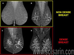
- Dense breast tissue also makes it harder for radiologists to see cancer on mammograms. Dense (fibrous and glandular) breast tissue looks white on a mammogram. Breast masses and cancers can also look white, so the dense tissue can make it harder to see them. In contrast, fatty tissue looks almost black on a mammogram, so it’s easier to see a tumor that looks white if most of the breast is fat tissue.
If I have dense breasts, do I still need a mammogram?
Yes. Most breast cancers can be seen on a mammogram even in women who have dense breast tissue, so it’s still important to get regular mammograms. Mammograms can help save women’s lives.
Even if you have a normal mammogram report, you should know how your breasts normally look and feel. Anytime there’s a change, you should report it to a health care provider right away.
Should I have any other screening tests if I have dense breast tissue?
At this time, experts do not agree what other tests, if any, should be done in addition to mammograms in women with dense breasts.
Digital breast tomosynthesis (3D mammography) can find some cancers not seen on regular (2D) mammograms. Some studies have suggested 3D mammography might be particularly helpful in women with dense breasts. It can be used as a screening test along with or instead of standard mammography, although it isn’t yet available at all imaging centers.
Studies have shown that breast ultrasound and possibly magnetic resonance imaging (MRI) can also help find some breast cancers that can’t be seen on mammograms. But ultrasound and MRI can also show more findings that are not cancer. This can lead to more tests and unnecessary biopsies.
Talk to your health care provider about whether you should consider any of these tests.
What should I do if I have dense breast tissue?
If your mammogram report says that you have dense breast tissue, talk with your health care provider about what this means for you. Be sure that your doctor or nurse knows if there’s anything in your medical history that increases your risk for breast cancer. To learn more about breast cancer risk factors, see Breast Cancer Risk and Prevention.
Any woman who’s already in a high-risk group (based on inherited gene mutations, a strong family history of breast cancer, or other factors) should have an MRI along with her yearly mammogram. To learn more about if you’re in a higher-risk group for breast cancer, see American Cancer Society Recommendations for the Early Detection of Breast Cancer.
What tests are recommended for breast cancer screening?
Most medical organizations recommend women with an average risk of breast cancer consider regular mammogram testing beginning at age 40 and consider repeating the screening annually.
Women with dense breasts, but no other risk factors for breast cancer, are considered to have a higher risk of breast cancer than average. They may benefit from annual breast cancer screening.
Dense breast tissue makes it more difficult to interpret a mammogram, since cancer and dense breast tissue both appear white on a mammogram. Very dense breasts may increase the risk that cancer won’t be detected on a mammogram.
Despite concerns about detecting cancer in dense breasts, mammograms are still effective screening tools. The most common type of mammogram — digital mammogram — saves images of your breasts as digital files instead of film and allows for more detailed analysis. This is more effective at finding cancer in dense breast tissue than older film mammogram technology.
Are other tests more effective?
There’s some evidence that additional tests may make it more likely that breast cancer is detected in dense breast tissue. But additional tests carry additional risks, and no additional testing method is proved to reduce the risk of dying of breast cancer.
You and your doctor may consider additional or supplemental testing based on your other risk factors and your personal preferences.
Supplemental tests for breast cancer screening may include:
- 3-D mammogram (breast tomosynthesis).
- Tomosynthesis uses X-rays to collect multiple images of the breast from several angles. The images are synthesized by a computer to form a 3-D image of the breast. Many mammogram centers are transitioning to incorporate 3-D mammograms as part of the standard mammogram technology.
-
Breast MRI.
- MRI uses magnets to create images of the breast. MRI doesn’t use radiation. Breast MRI is recommended for women with a very high risk of breast cancer, such as those with genetic mutations that increase the risk of cancer.
- Breast ultrasound.
- Ultrasound uses sound waves to analyze tissue. A diagnostic ultrasound is commonly used to investigate areas of concern discovered on a mammogram.
- Molecular breast imaging (MBI)
- . MBI, also known as breast-specific gamma imaging, uses a special camera (gamma camera) that records the activity of a radioactive tracer. The tracer is injected into a vein in your arm. Normal tissue and cancerous tissue react differently to the tracer, which can be seen in the images produced by the gamma camera. MBI is performed every other year in addition to an annual mammogram.
Every test has pros and cons. While each test is proved to find more breast cancers than a mammogram, none of these newer imaging tests is proved to reduce the risk of dying of breast cancer, as has been done with the standard film mammogram.