largest synovial joint
Hi,welcome to solsarin site, today we want to talk about “largest synovial joint” ,thank you for choosing us.
Table of contents
1.what is the largest synovial joint in body?
2.What is the largest and most complex synovial joint?
largest synovial joint
A synovial joint is a connection between two bones which consists of a cartilage lined cavity filled with fluid. The knee joint is the biggest synovial joint in the human body. At the junction of the knee, the large femur bone of the thigh connects to both the tibia and fibula of the leg, as well as to the patella or knee cap. So, the correct answer is ‘Knee joint’.
knee, hinge joint that is formed by the meeting of the thigh bone (femur) and the larger bone (tibia) of the lower leg. The knee is the largest joint in the body and has to sustain the greatest stresses, since it supports the entire weight of the body above it.
 Knee joint
 Knee joint
The three joints in the body (Histologically) are fibrous, cartilaginous, and synovial
Joint
A joint is the part of the body where two or more bones meet to allow movement. Generally speaking, the greater the range of movement, the higher the risk of injury because the strength of the joint is reduced. The six types of freely movable joint include ball and socket, saddle, hinge, condyloid, pivot and gliding.
Synovial joints
Synovial joints are the most common type of joint in the body. These joints are termed diarthroses, meaning they are freely mobile.
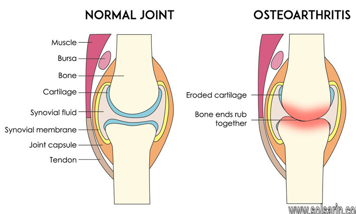
A key structural characteristic for a synovial joint that is not seen at fibrous or cartilaginous joints is the presence of a joint cavity. The joint cavity contains synovial fluid, secreted by the synovial membrane (synovium), which lines the articular capsule. This fluid-filled space is the site at which the articulating surfaces of the bones contact each other. Hyaline cartilage forms the articular cartilage, covering the entire articulating surface of each bone. The articular cartilage and the synovial membrane are continuous. A few synovial joints of the body have a fibrocartilage structure located between the articulating bones. This is called an articular disc, which is generally small and oval-shaped, or a meniscus, which is larger and C-shaped.
synovial fluid
Synovial fluid, also known as joint fluid, is a thick liquid located between your joints. The fluid cushions the ends of bones and reduces friction when you move your joints. A synovial fluid analysis is a group of tests that checks for disorders that affect the joints.
Synovial fluid is the thick liquid that lubricates your joints and keeps them moving smoothly. It’s on all of your joints, including in your knees, shoulders, hips, hands, and feet.
Joint conditions like arthritis, gout, infections, and bleeding disorders can change how your synovial fluid looks and feels. A sample of this fluid taken during a procedure called an arthrocentesis can help your doctor figure out what’s causing your symptoms.
arthritis
Arthritis is the swelling and tenderness of one or more joints. The main symptoms of arthritis are joint pain and stiffness, which typically worsen with age. The most common types of arthritis are osteoarthritis and rheumatoid arthritis.
gout
Gout is a common and complex form of arthritis that can affect anyone. It’s characterized by sudden, severe attacks of pain, swelling, redness and tenderness in one or more joints, most often in the big toe.
bleeding disorders
Bleeding disorders are a group of conditions in which there is a problem with the body’s blood clotting process. These disorders can lead to heavy and prolonged bleeding after an injury. Bleeding can also begin on its own. Specific bleeding disorders include: Acquired platelet function defects.
synovium
The term synovium refers to the soft tissue lining the spaces of diarthrodial joints, tendon sheaths and bursae. It includes the continuous surface layer of cells (intima) and the underlying tissue (subintima).
The synovium has a rich network of sympathetic and sensory nerves. The former, which are myelinated and detected with the antibody against S-100 protein, terminate close to blood vessels, where they regulate vascular tone (Figure 2-6C through E). Sensory nerves respond to proprioception and pain via large myelinated nerve fibers and via small (<5 µm) unmyelinated or myelinated fibers with unmyelinated free nerve ends (nociceptors). The latter are immunoreactive in the synovium for neuropeptides, including substance P, calcitonin gene–related peptide, and vasoactive intestinal peptides.
cartilage
Cartilage is an important structural component of the body. It is a firm tissue but is softer and much more flexible than bone. Cartilage is a connective tissue found in many areas of the body including: Joints between bones e.g. the elbows, knees and ankles. Ends of the ribs.
Cartilage is a unique tissue type because it doesn’t have blood vessels or nerves. Instead, cartilage cells (known as chondrocytes) are found in a gel-like “matrix” that provides nourishment to the cells. Cartilage has a unique structure that makes it a strong but flexible tissue.
meniscus
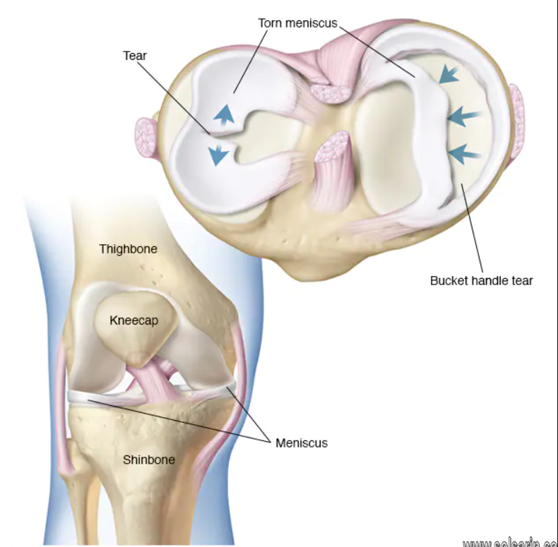
The meniscus is a C-shaped piece of tough, rubbery cartilage that acts as a shock absorber between your shinbone and thighbone. It can be torn if you suddenly twist your knee while bearing weight on it. A torn meniscus is one of the most common knee injuries.
What is a meniscus tear?
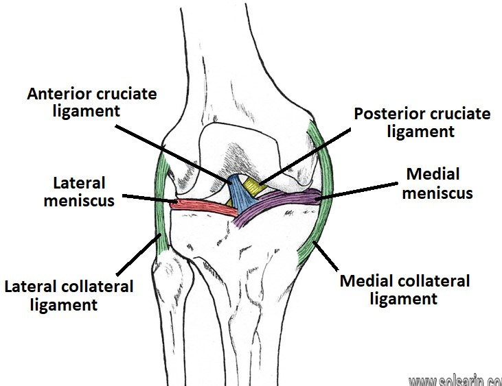
The menisci sit between the tibia (lower leg bone) and the femur (thigh bone) and protect the lower part of the leg from the shock created by our body weight. The medial meniscus sits on the inside of the knee and the lateral meniscus sits on the outside of the knee.
Meniscus tears usually take place when an athlete twists or turns their upper leg while their foot is planted and their knee is bent.
Occasionally menisci can develop as a block or disk shape, which is called a discoid meniscus. A discoid meniscus is more likely to tear and commonly presents in childhood.