mild diaphragmatic attenuation
Hello dear friends, thank you for choosing us. In this post on the solsarin site, we will talk about “mild diaphragmatic attenuation”.
Stay with us.
Thank you for your choice.
What does diaphragmatic attenuation mean?
In respect to this, what is breast attenuation?
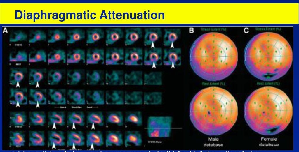
BACKGROUND: Breast attenuation artifact is well known for reducing the accuracy of myocardial perfusion imaging in women. The normals were further divided into those with breast attenuation (defined as mean anterior counts <70% maximum) and those without.
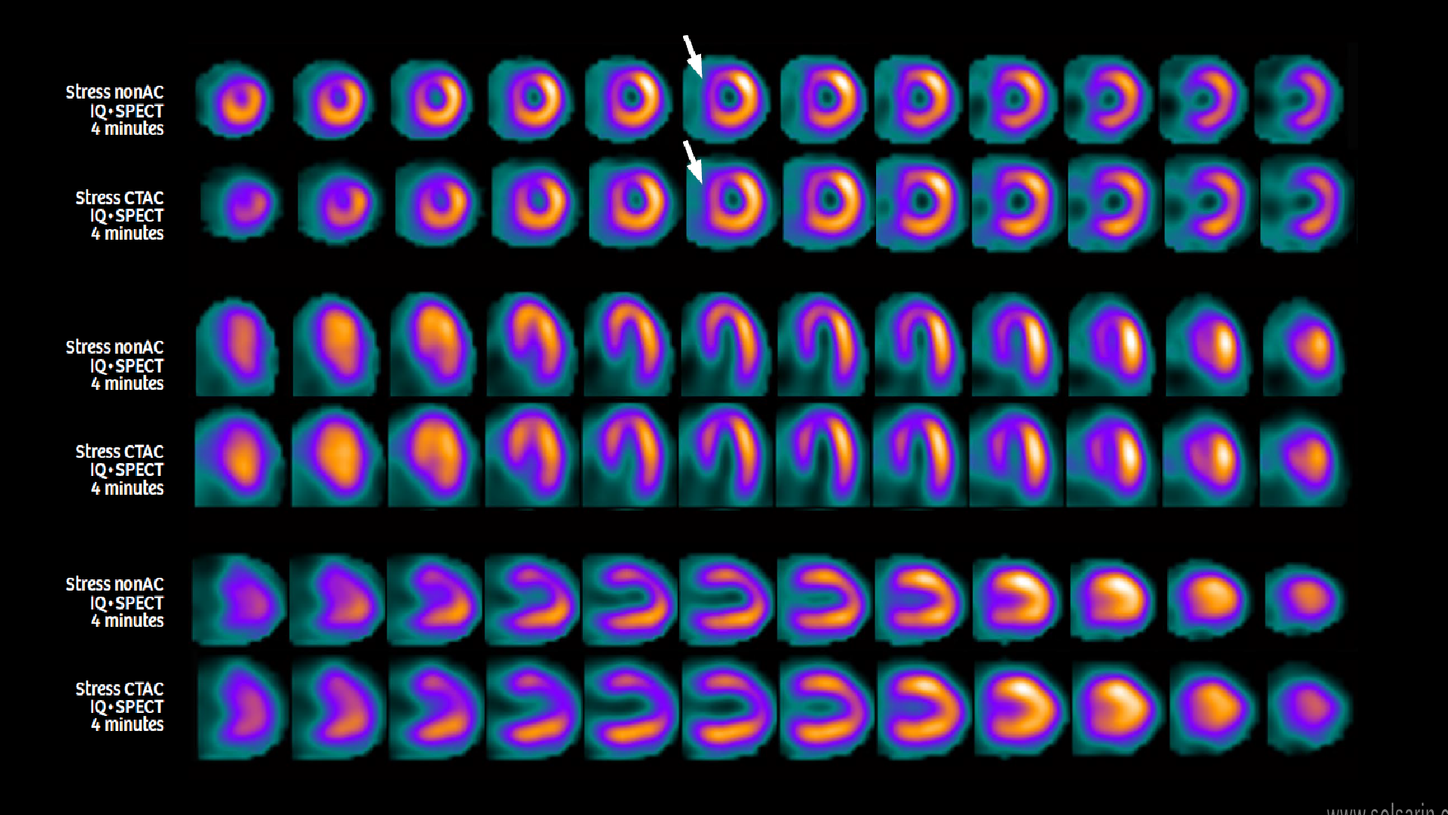
Also Know, what is gut attenuation? Attenuation correction is a mechanism that removes soft tissue artifacts from SPECT images. Attenuation artifacts vary among patients, but the most common corrections are to artifacts associated with breast attenuation in women and diaphragmatic attenuation in men.
Considering this, what causes attenuation artifact?
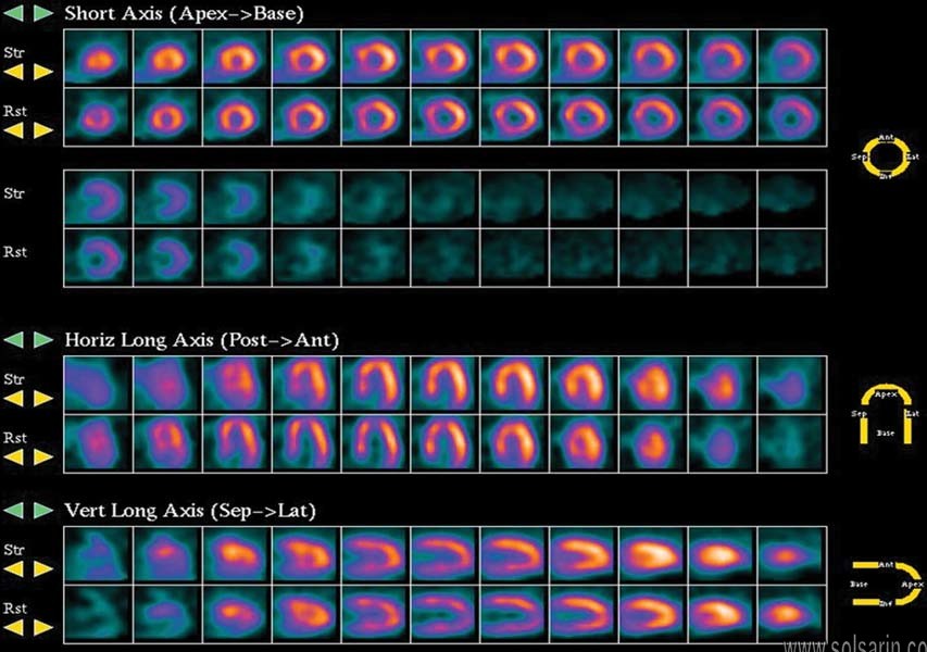
“Attenuation artifact” observed in a nuclear stress test is due to the reduction in the intensity / strength of signal when it travels through various body tissues of different densities, such as breast tissues, chest wall, and organs under the diaphragm.
What does soft tissue attenuation mean?
After a stress test, your physician may mention that you have “soft tissue artifact,” or “soft tissue attenuation.” What this means is that soft tissue, such as breast tissue, is showing up on the image created by the stress test.
Avoidance of falsely positive results depends on distinguishing reality from artifact, in turn depending on images of highest quality. In radionuclide cardiac imaging, an inferior wall artifactual defect, so called “diaphragmatic attenuation”, is particularly common and vexing. Despite the historically held view, analysis and review of the literature suggest the defect is likely not diaphragmatic but rather primarily due to attenuation by nearby stomach wall.
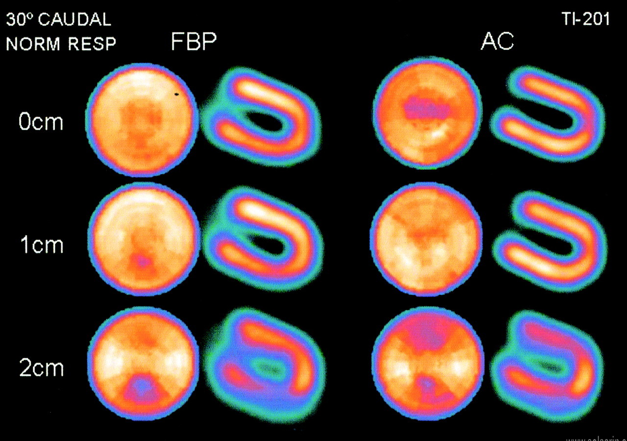
The explanation is based on gravity and anatomy. With this improved understanding, effervescent granules were given as a clinical, nonresearch measure to nine patients during myocardial scanning. It was observed that two-thirds demonstrated moderate or marked lessening of attenuation. An additional benefit is lessening of artifact by extracardiac activity. These benefits may also apply to other sorts of cardiac radionuclide imaging. The significance of this new imaging method is discussed and various avenues of research are proposed.
What is an artifact in medical terms?
What is attenuation correction in pet?
What is a fixed apical defect?
What is a perfusion defect?
What is ultrasound artifact?
What is abnormal myocardial perfusion?
What is an artifact on a stress test?
What does artifact mean on ECG?
What is network attenuation?