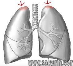
uppermost portion of the lung
Hello dear readers. In this post on Solsarin we are going to talk about ”uppermost portion of the lung “. Continue reading to find the answer.please write your comment, Thank you for your attention.
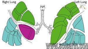
Bronchial tree
The lungs begin at the bottom of your trachea (windpie). The trachea is a tube that carries the air in and out of your lungs. Each lung has a tube called a bronchus that connects to the trachea. The trachea and bronchi airways form an upside-down “Y” in your chest. This “Y” is often called the bronchial tree.
The bronchi branch off into smaller bronchi and even smaller tubes called bronchioles. Like the branches of a tree, these tiny tubes stretch out into every part of your lungs. Some of them are so tiny that they have the thickness of a hair. You have almost 30,000 bronchioles in each lung.
Each bronchiole tube ends with a cluster of small air sacs called alveoli (individually referred to as alveolus). They look like tiny grape bunches or very tiny balloons. There are about 600 million alveoli in your lungs. The small bubble shapes of the alveoli give your lungs a surprising amount of surface area — equivalent to the size of a tennis court. This means there’s plenty of room for vital oxygen to pass into your body.
SUMMARYEach lung is divided into lobes. The bronchial tree running through your lungs is made up of the windpipe, bronchi, bronchioles, and alveoli.
The respiratory system
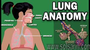
The lungs are the main part of the respiratory system. This system is divided into the upper respiratory tract and the lower respiratory tract.
The upper respiratory tract includes the:
- Mouth and nose. Air enters and leaves the lungs through the mouth and nostrils of the nose.
- Nasal cavity. Air passes from the nose into the nasal cavity, and then the lungs.
- Throat (pharynx). Air from the mouth is sent to the lungs via the throat.
- Voice box (larynx). This part of the throat helps air to pass into the lungs and keeps out food and drink.
The lower respiratory tract is made up of the:
- lungs
- trachea (windpipe)
- bronchi
- bronchioles
- alveoli
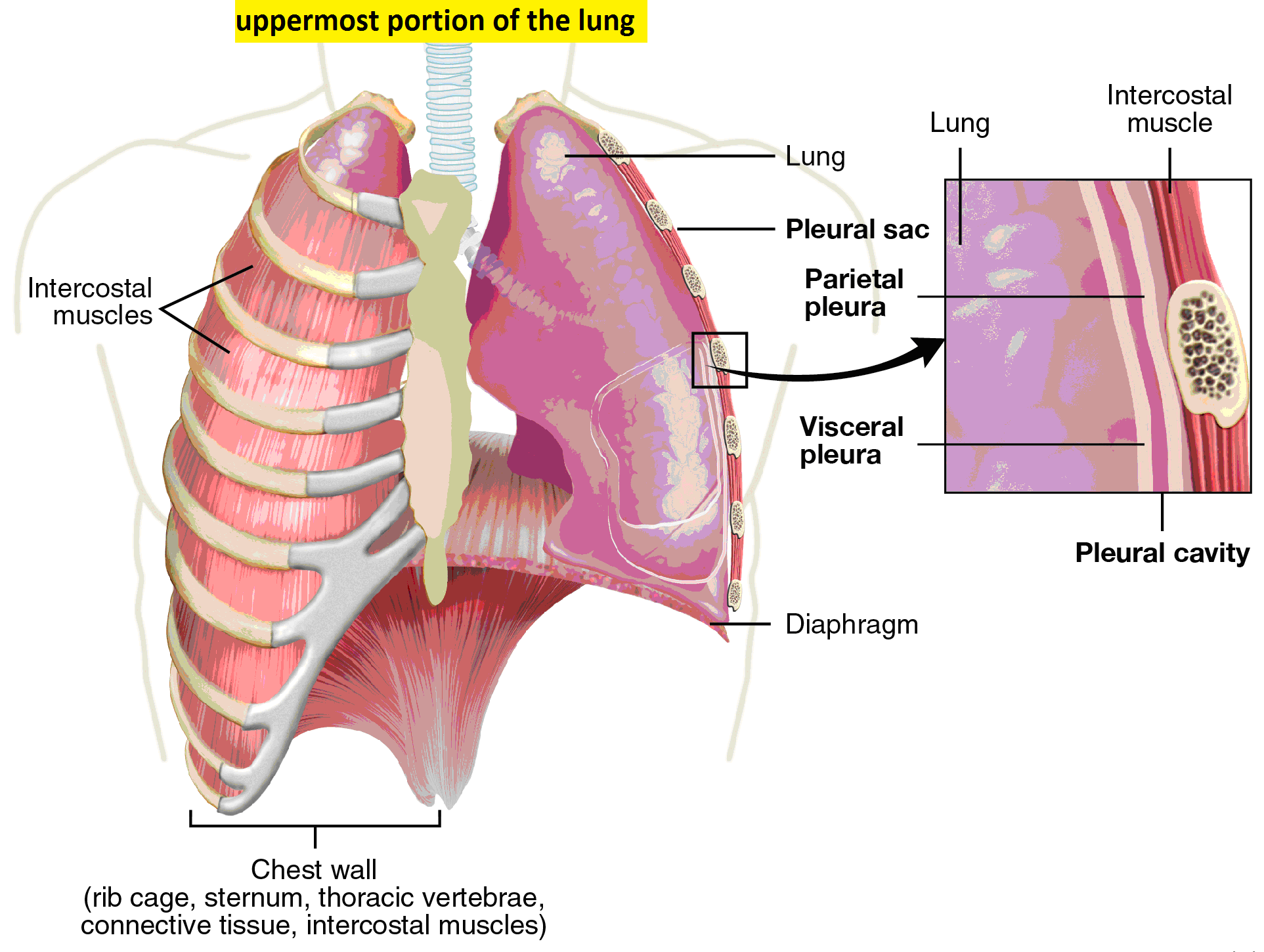
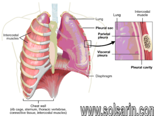
Other parts of the respiratory system help your lungs to expand and contract as you breathe. These include the ribs around the lungs and the dome-shaped diaphragm muscle below them.
ADVERTISEMENT
The Ultimate COPD Survival Guide
Are you experiencing chronic shortness of breath or a nagging cough? You’ve come to the right place. Here’s everything you need to know to manage your COPD symptoms.

The lungs are surrounded by your sternum (chest bone) and ribcage on the front and the vertebrae (backbones) on the back. This bony cage helps to protect the lungs and other organs in your chest.
How your lungs work
WHAT IS RESPIRATION?Respiration is made up of two phases called inspiration and expiration: You inhale (breathe in) oxygen during inspiration. You exhale (breathe out) carbon dioxide during expiration.
The pathway of a breath
When you breathe, air enters through your mouth and nose and travels:
- down the throat into the trachea
- into the lungs through the right and left main bronchi
Each alveolus is covered by a net of tiny blood vessels called capillaries. Oxygen and carbon dioxide exchange happens here. Your heart sends deoxygenated blood to the lungs. This is blood that is carrying carbon dioxide rather than oxygen.
As the blood passes through the tiny, thin-walled capillaries, they get oxygen from the alveoli. They return carbon dioxide through the thin walls to the alveoli.
The oxygen-rich blood from your lungs is sent back to your heart, where it’s pumped to your entire body. The carbon dioxide is breathed out of the lungs and alveoli through your mouth and nose.
How your lungs stay healthy
The alveoli stay partly inflated like a balloon even when you exhale air. Your lungs make a fluid called surfactant to help them stay open. Surfactant also contains fatty proteins that help keep the lungs healthy.
Your lungs are self-cleaning.
They make mucus to trap germs and particles. The mucus is then swept up by cilia, small hairs that line the airways. Normally, you swallow this mucus without noticing. If you have a respiratory illness, your lungs may make too much mucus.
The alveoli also contain immune cells called macrophages. These cells “eat” germs and irritants before they can cause an infection in your lungs.
Lung disorders and illnesses
A respiratory disorder may be temporary or chronic (long term). Some types may lead to, or be a sign of, lung disease. Common lung conditions include:
Asthma
Asthma is the most common chronic lung condition. Allergic asthma typically begins in childhood. Asthma attacks happen when the airways tighten and narrow, slowing down air flow. The lungs also become swollen and inflamed.
Asthma can be triggered by an allergic reaction, pollution, exercise, other respiratory illness, and cold air.
Bronchitis
This chest infection happens in the main airways, the bronchi. It may be due to a viral or bacterial infection.
Acute bronchitis happens suddenly and can sometimes spread into the lungs from an upper respiratory tract infection, such as a common cold.
Chronic obstructive pulmonary disease (COPD)
This condition is also known as chronic bronchitis or emphysema. COPD gets worse over time. It may be caused by smoking, air pollution, chemicals, or a genetic condition.
COPD often leads to disability and is the fourth most common cause of deathTrusted Source in the U.S.
Pneumonia
This is a chest infection deep in the bronchioles and alveoli. Pus and mucus can build up, and the lungs may swell. This makes it difficult to breathe. Pneumonia can happen to anyone. Young children, the elderly, smokers, and people who are ill are at higher risk.
Tuberculosis (TB)
This bacterial infection is spread through air droplets from coughs and sneezes. It’s difficult to become infected. Tuberculosis can be serious and lead to lung scarring. It may also stay in the body without causing symptoms or spread to other parts of the body.
Causes of lung disorders and illnesses
Respiratory or lung disorders can make it difficult to breathe. They are a common reason for doctor visits in most countries.
You can get a respiratory illness due to:
- bacteria
- viruses
- mold (fungus)
- polluted air
- chemicals
- stagnant indoor air
- cigarette, tobacco, or shisha smoke
- secondhand smoke
- allergies, such as:
- pollen
- dust
- food allergens (dairy, nuts, seafood, etc.)
- pet dander and fur
- insect waste (such as from dust mites)
Symptoms to see a doctor about
See your doctor if you experience severe lung symptoms. According to the American Lung Association, warning signs of lung disease include:
- chronic cough that lasts for a month or longer
- shortness of breath after little or no exertion
- wheezing or noisy breathing
- chronic mucus or sputum in your lungs that lasts for a month or longer
- chronic chest pain that lasts for a month or longer
- coughing up blood
Lung function tests
If you have a respiratory disorder, you may need tests to see how well your lungs are working. They also help to diagnose chronic lung illness. Some of these tests are routine for people with chronic illness such as asthma. Common lung function tests and scans include:
- Arterial blood gas tests. This test measures blood oxygen levels. You will need a blood test, which requires blood to be drawn. The blood sample is sent to a lab to measure the amount of oxygen and carbon dioxide in it.
- Blood test. A blood test checks for a bacterial or viral infection. It also checks your white blood cell count. A high count may mean that you have an infection.
- Chest X-ray. This helps your doctor to see how healthy your lungs are. An X-ray will show areas of the lung that are clogged up or scarred. Your doctor may also recommend other types of lung scans.
- Exhaled nitric oxide test. Nitric oxide helps to relax blood vessels and airways. This increases blood flow to your lungs, improving oxygen levels. Nitric oxide levels can show if certain medications will treat your asthma. You will need to breathe into a tube for this test.