Which microscope is used to see internal structures of cells in a natural state?
Hey guys! We return with an amazing topic about science and health. This is “Which microscope is used to see internal structures of cells in a natural state?”. As always, read the text, share it to your friends and comment us in solsarin.
Which microscope is used to see internal structures of cells in a natural state?
Electron microscopes
Electron microscopes can be used to examine not just whole cells, but also the subcellular structures and compartments within them.
Microscopy
Introduction
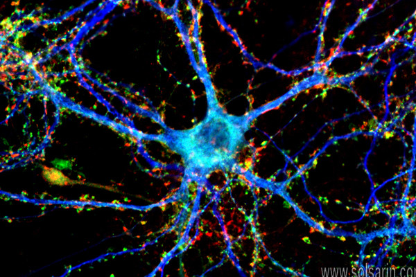
If you meet some cell biologists and get them talking about what they enjoy most in their work, you may find it comes down to one thing: secretly, they’re all microscope freaks. At the end of the day, what they really love is the chance to sit in a small, dark room for hours on end, communing with their favorite cell type through the lens of a beautiful microscope.
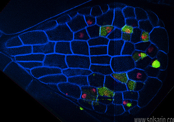
That may seem odd, but the truth is, cells can be pretty gorgeous, like living stained glass. One of my favorite examples of this is the picture below, which shows cells in a very young leaf of thale cress, a small flowering plant related to mustard.
You could find cells just as intricately patterned and beautifully formed in any plant you looked at – from the rose in your backyard, to the grass growing up through the sidewalk, to the carrots you ate for a snack.
Let’s not limit it to plants, either: exquisite layers of cells can be found in your skin, in an insect’s wing, and in just about any other living tissue you choose to look at. We, and the world around us, are cathedrals made of cells. We just need some microscopy to appreciate it.
Microscopes and lenses
Although cells vary in size, they’re generally quite small. For instance, the diameter of a typical human red blood cell is about eight micrometers (0.008 millimeters). To give you some context, the head of a pin is about one millimeter in diameter, so about 125 red blood cells could be lined up in a row across the head of a pin.
With a few exceptions, individual cells cannot be seen with the naked eye, so scientists must instead use microscopes (micro– = “small”; –scope = “to look at”) to study them. A microscope is an instrument that magnifies objects otherwise too small to be seen, producing an image in which the object appears larger. Most photographs of cells are taken using a microscope, and these pictures can also be called micrographs.
From the definition above, it might sound like a microscope is just a kind of magnifying glass. In fact, magnifying glasses do qualify as microscopes; since they have just one lens, they are called simple microscopes.
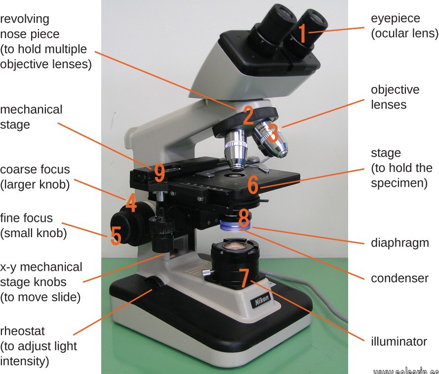
The fancier instruments that we typically think of as microscopes are compound microscopes, meaning that they have multiple lenses. Because of the way these lenses are arranging, they can bend light to produce a much more magnified image than that of a magnifying glass.
In a compound microscope with two lenses, the arrangement of the lenses has an interesting consequence: the orientation of the image you see is flipped in relation to the actual object you’re examining.
For example, if you were looking at a piece of newsprint with the letter “e” on it, the image you saw through the microscope would be “ə.” start superscript, 1, end superscript More complex compound microscopes may not produce an inverted image because they include an additional lens that “re-inverts” the image back to its normal state.
What separates a basic microscope from a powerful machine used in a research lab? Two parameters are especially important in microscopy: magnification and resolution.
-
Magnification is a measure of how much larger a microscope (or set of lenses within a microscope) causes an object to appear. For instance, the light microscopes typically used in high schools and colleges magnify up to about 400 times actual size. So, something that was 1 mm wide in real life would be 400 mm wide in the microscope image.
-
The resolution of a microscope or lens is the smallest distance by which two points can be separated and still be distinguished as separate objects. The smaller this value, the higher the resolving power of the microscope and the better the clarity and detail of the image. If two bacterial cells were very close together on a slide, they might look like a single, blurry dot on a microscope with low resolving power, but could be told apart as separate on a microscope with high resolving power.
Both magnification and resolution are important if you want a clear picture of something very tiny. For example, if a microscope has high magnification but low resolution, all you’ll get is a bigger version of a blurry image. Different types of microscopes differ in their magnification and resolution.
Electron microscopes
Some cutting-edge types of light microscopy (beyond the techniques we discussed above) can produce very high-resolution images. However, if you want to see something very tiny at very high resolution, you may want to use a different, tried-and-true technique: electron microscopy.
Electron microscopes differ from light microscopes in that they produce an image of a specimen by using a beam of electrons rather than a beam of light. Electrons have much a shorter wavelength than visible light; and this allows electron microscopes to produce higher-resolution images than standard light microscopes.
Electron microscopes can be used to examine not just whole cells, but also the subcellular structures and compartments within them.
One limitation; however, is that electron microscopy samples must be placed under vacuum in electron microscopy; (and typically are prepared via an extensive fixation process). This means that live cells cannot be imaged.