pertaining to under the collarbone
Hello dear friends, thank you for choosing us. In this post on the solsarin site, we will talk about “pertaining to under the collarbone”.
Stay with us.
Thank you for your choice.

pertaining to under the collarbone
Medical Definition of Collarbone
Collarbone: A horizontal bone above the first rib that makes up the front part of the shoulder.
One end of the collarbone connects to the sternum, forming one side of the sternoclavicular joint.
The other end of the collarbone connects to the scapula, there forming one side of the acromioclavicular joint.
sports injuries
Failing to warm up increases the risk of sports injuries. Bruises, strains, sprains, tears, and broken bones can result from sports injuries.
Soft tissues like muscles, ligaments, tendons, fascia, and bursae may be affected. Injuries may range from mild to severe.
Pulled Muscle
- pain,
- swelling,
- weakness, and
- difficulty or inability to use the muscle.
Muscles in the quadriceps, the calves, hamstrings, groin, low back, and shoulder are the most common sites for pulled muscles. Minor muscle strains resolve with RICE — Rest, Ice.
Compression, and Elevation. Nonsteroidal anti-inflammatory drugs (NSAIDs) may help manage pain and swelling as well. More serious muscle strains require evaluation and treatment by a doctor.
Torn ACL
The anterior cruciate ligament (ACL) helps hold the knee joint together and provides stability.
. Pain, swelling, and loss of range of motion are symptoms of a torn ACL.It may be difficult to walk. A torn ACL needs to be reconstructed surgically, usually using a graft from another ligament in the patient’s own body.
Significant rehabilitation is necessary to restore the strength and function of the knee joint after surgery. Depending on the age, health status.
and desired activity level of the patient, some may not elect to have surgery.
Torn MCL
The medial collateral ligament (MCL) connects the upper leg bone (femur) to the larger bone of the lower leg (tibia). It is located on the inner side of the knee.
A torn MCL results in pain, swelling, and instability of the joint. The condition is often treated with ice, bracing, and physical therapy.
If other structures in the knee are injured or if the torn MCL is severe, surgery may be recommended.
stabbing pain
Shin splint symptoms are throbbing, aching, or stabbing pain on the insides of the lower leg.
Pain occurs when muscles and tendons around the tibia (the larger of the two lower leg bones).
become inflamed. Stretching, resting, and applying ice can help relieve shin splints.
Nonsteroidal anti-inflammatory drugs (NSAIDs) can reduce pain and swelling.
Bandaging the area may help prevent swelling. Flat feet increase the risk of shin splints.
Orthotics and proper athletic shoes may offer support and decrease the risk of shin splints.
Stress Fracture
and a bone absorbs the pressure, resulting in a break. feet.
. Stress fractures cause pain with activity. Rest is prescribed to allow a stress fracture to heal.
Sometimes a special shoe or a brace helps decrease stress on the bone, which facilitates healing.
Plantar Fasciitis
The plantar fascia is a ligament that connects the heel to the front of the foot, supporting the arch. Plantar fasciitis is inflammation of this ligament.
It causes heel pain often felt the first thing in the morning after getting out of bed or after being active.
Stress and strain on the feet increases the risk of plantar fasciitis.
Obesity, tight calf muscles, repetitive use, high arches, and new athletic activities are all risk factors for this condition. Plantar fasciitis is treated with rest.
ice, nonsteroidal anti-inflammatory drugs (NSAIDs), and special stretching exercises.
insoles may provide relief. Wearing splints at night may help decrease pain.
More severe cases of plantar fasciitis may be treated with cortisone injections, physical therapy, and/or surgery.
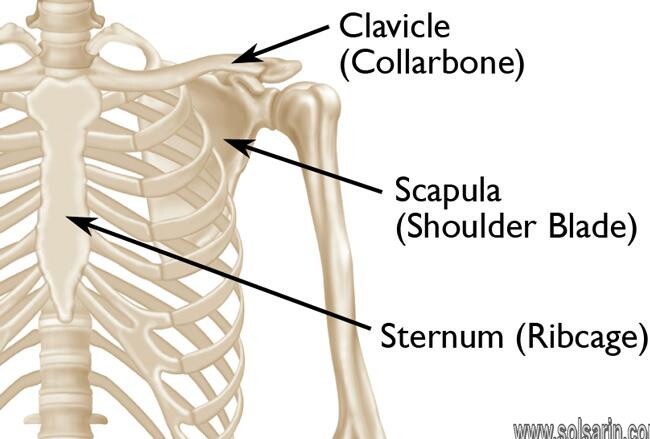
The clavicle
The clavicle (collarbone) extends between the manubrium of the sternum and the acromion of the scapula.
It is classed as a long bone and can be palpated along its length.
In thin individuals, it is visible under the skin. The clavicle has three main functions:
- Attaches the upper limb to the trunk as part of the ‘shoulder girdle’.
- Protects the underlying neurovascular structures supplying the upper limb.
- Transmits force from the upper limb to the axial skeleton.
In this article, we shall look at the anatomy of the clavicle – its bony landmarks and clinical correlations.
Bony Landmarks and Articulations
The clavicle is a slender bone with an ‘S’ shape. Facing forward.
the medial aspect is convex, and the lateral aspect concave.
It can be divided into a sternal end, a shaft and an acromial end.
Sternal (medial) End
The sternal end contains a large facet .
– for articulation with the manubrium of the sternum at the sternoclavicular joint
The inferior surface of the sternal end is marked by a rough oval depression for the costoclavicular ligament (a ligament of the SC joint).
Shaft
The shaft of the clavicle acts a point of origin and attachment for several muscles – deltoid, trapezius, subclavius, pectoralis major, sternocleidomastoid and sternohyoid
Acromial (lateral) End
The acromial end houses a small facet for articulation with the acromion of the scapula at the acromioclavicular joint.
The clavicle (collarbone) extends between the manubrium of the sternum and the acromion of the scapula.
It is classed as a long bone
and can be palpated along its length. In thin individuals, it is visible under the skin. The clavicle has three main functions:
- Attaches the upper limb to the trunk as part of the ‘shoulder girdle’.
- Protects the underlying neurovascular structures supplying the upper limb.
- Transmits force from the upper limb to the axial skeleton.
In this article, we shall look at the anatomy of the clavicle – its bony landmarks and clinical correlations.
Fig 1 – The anatomical position of the clavicle
Bony Landmarks and Articulations
The clavicle is a slender bone with an ‘S’ shape. Facing forward, the medial aspect is convex, and the lateral aspect concave.
Sternal (medial) End
The sternal end contains a large facet – for articulation with the manubrium of the sternum at the sternoclavicular joint.
Shaft
The shaft of the clavicle acts a point of origin and attachment for several muscles .
– deltoid, trapezius, subclavius, pectoralis major, sternocleidomastoid and sternohyoid
Acromial (lateral) End
The acromial end houses a small facet for articulation with the acromion of the scapula at the acromioclavicular joint.
- Conoid tubercle – attachment point of the conoid ligament, the medial part of the coracoclavicular ligament.
- Trapezoid line – attachment point of the trapezoid ligament, the lateral part of the coracoclavicular ligament.
The coracoclavicular ligament is a very strong structure, effectively suspending the weight of the upper limb from the clavicle.
Overview
Your collarbone connects the upper part of your breastbone to your shoulder blade.
Common causes of a broken collarbone include falls, sports injuries and trauma from traffic accidents.
When to see a doctor
If you notice signs or symptoms of a broken collarbone in you or your child.
or if there’s enough pain to prevent normal use, see a doctor right away. Delays in diagnosis and treatment can lead to poor healing.
Causes
Common causes of a broken collarbone include:
- Falls, such as falling onto your shoulder or onto your outstretched hand.
- Sports injuries, such as a direct blow to your shoulder on the field, rink or court.
- Vehicle trauma from a car, motorcycle or bike accident.
- Birth injury from passing through the birth canal.
Risk factors
Your collarbone doesn’t harden completely until about age 20.
This puts children and teenagers at higher risk of a broken collarbone.
The risk decreases after age 20, but then rises again in older people as bone strength decreases with age.
Complications
Most broken collarbones heal without difficulty. Complications, when they occur, might include:
Clavicle
The clavicle, or collarbone, is a slender, S-shaped long bone approximately 6 inches (15 cm) long[1] .
that serves as a strut between the shoulder blade and the sternum (breastbone). There are two clavicles, one on the left and one on the right.
RESOURCE: WIKIPEDIA