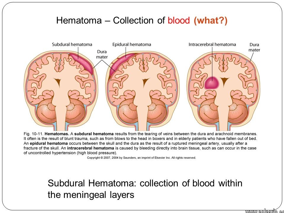
collection of blood within the meningeal layers
Hi, welcome to solsarin site, in this post we want to talk about“collection of blood within the meningeal layers”,
stay with us.
collection of blood within the meningeal layers
The blood supply of the meninges generally concerns the blood supply of the outer layer of dura mater rather than the inner layer of dura mater, arachnoid or pia mater which do not require a large blood supply. There are several arteries that supply the dura in the anterior, middle, and posterior cranial fossae.
Blood supply
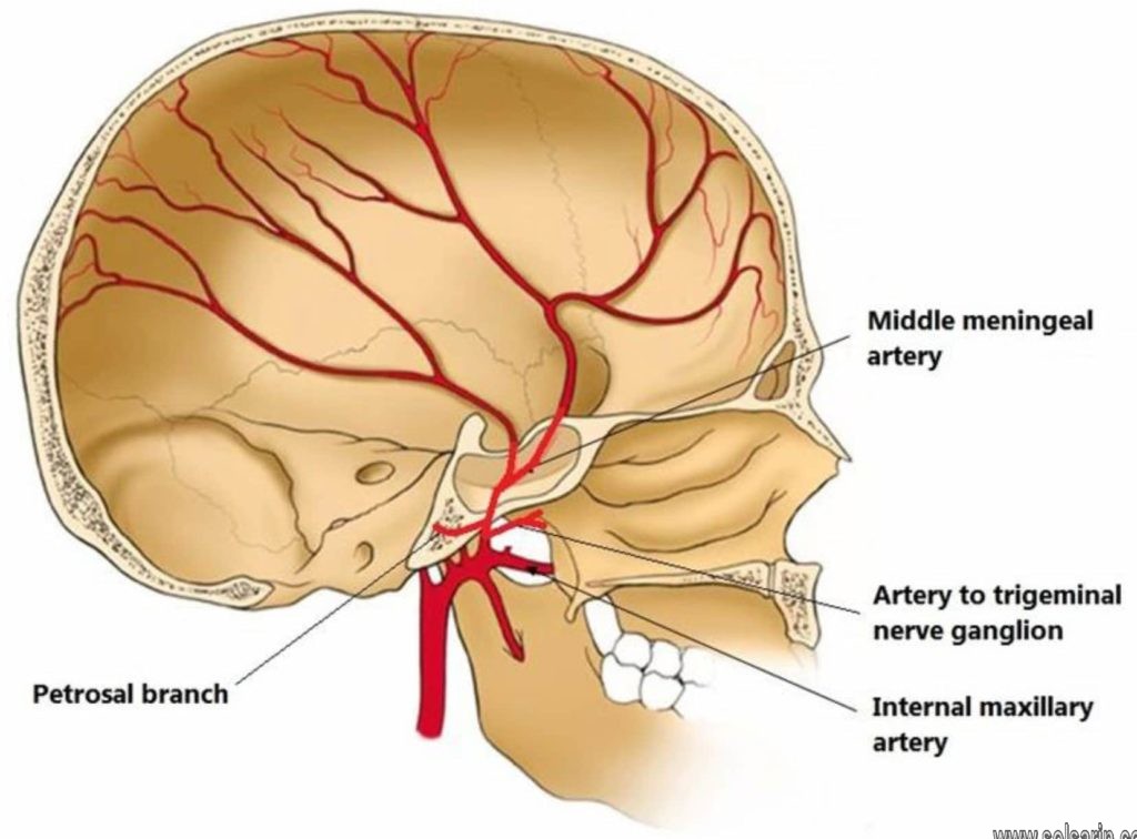
The cranial meninges primarily receive blood from by the middle meningeal artery (MMA), which is a branch of the internal carotid artery, which progresses up the neck. On each side, the MMA enters the skull through an opening in its side called the foramen spinosum and continues through the epidural space.
At the same opening—and running alongside the MMA—is the meningeal branch of the mandibular nerve. Its two branches transmit signaling between the outermost meningeal layer and the brain, with its anterior branch communicating with the meningeal branch of the maxillary nerve.
Blood supply for the spinal meninges comes from a single anterior artery, as well as two paired posterior spinal arteries.2 Branches of the vertebral arteries, they arise at the base of the neck and take an upward course.
Anterior cranial fossa
- meningeal branches of
- anterior ethmoidal artery
- posterior ethmoidal artery
- ophthalmic artery
- frontal branch of the middle meningeal artery (which enters the middle cranial fossa via the foramen spinosum)
Middle cranial fossa
- frontal and parietal branches of the middle meningeal artery
- accessory meningeal artery
- ascending pharyngeal artery
- branches directly from the internal carotid artery
The supratentorial dura mater is primarily supplied by the middle meningeal artery. This is a branch of the maxillary artery, which, despite its name, primarily supplies the calvarium rather than the meninges . Clinical importance is given to this artery due to its location in the extradural space and the proximity of its anterior division to the pterion, making it susceptible to damage in head injuries.
Posterior cranial fossa
- vertebral arteries
- occipital arteries
- ascending pharyngeal arteries
Related pathology
- extradural hemorrhage
Meninges
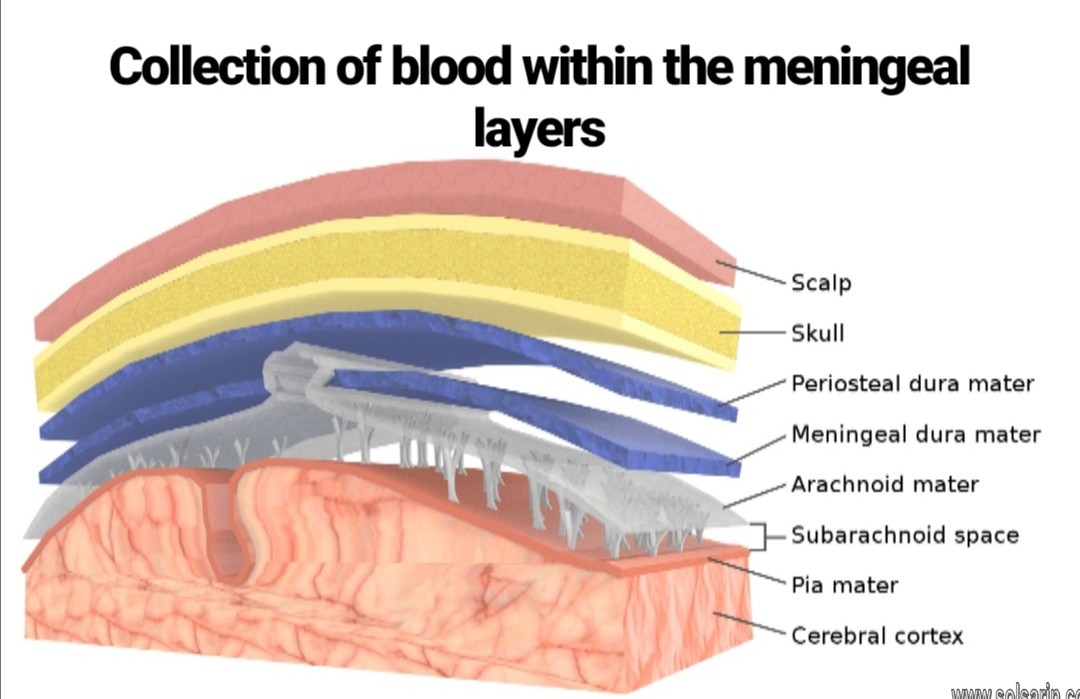
There are three layers of meninges around the brain and spinal cord. The outer layer, the dura mater, is tough, white fibrous connective tissue. The middle layer of meninges is arachnoid, a thin layer resembling a cobweb with numerous threadlike strands attaching it to the innermost layer. The space under the arachnoid, the subarachnoid space, is filled with cerebrospinal fluid and contains blood vessels. The pia mater is the innermost layer of meninges. This thin, delicate membrane is tightly bound to the surface of the brain and spinal cord and cannot be dissected away without damaging the surface.
Meningiomas are tumors of the nerve tissue covering the brain and spinal cord. Although meningiomas are unlikely to spread, physicians often treat them as though they were malignant because symptoms may develop when a tumor applies pressure to the brain.
What are meninges?
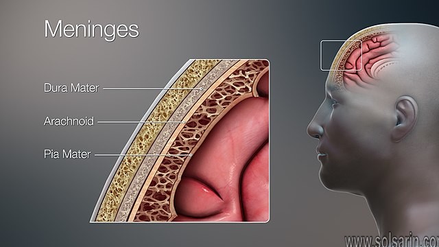
Meninges are three layers of membranes that cover and protect your brain and spinal cord (your central nervous system [CNS]). They’re known as:
- Dura mater: This is the outer layer, closest to your skull.
- Arachnoid mater: This is the middle layer.
- Pia mater: This is the inner layer, closest to your brain tissue.
Together, the arachnoid mater and pia mater are called leptomeninges.
There are three spaces within the meninges:
- The epidural space is a space between your skull and dura mater and the dura mater of your spinal cord and the bones of your vertebral column. Analgesics (pain medicine) and anesthesia are sometimes injected into this space along your spine. The spinal cord ends between the first and second lumbar vertebra in the middle of your back, at which point, only cerebrospinal fluid is present. This is the site where a lumbar puncture (“spinal tap”) is performed.
- The subdural space is a space between your dura mater and your arachnoid mater. Under normal conditions, this space isn’t a space, but can be opened if there’s trauma to your brain (such as a brain bleed) or other medical condition.
- The subarachnoid space is a space between your arachnoid mater and pia mater. It’s filled with cerebrospinal fluid. Cerebrospinal fluid cushions and protects your brain and spinal cord.
Where are the meninges?
The term meninges comes from the Greek for “membrane” and refers to the three membranes that surround the brain and spinal cord. The membrane layers (discussed in detail below) from the outside in are the: dura mater, arachnoid mater, and pia mater. Their positioning around the brain can be seen in the image to the right.
Meninges Layers
The meninges can be generally separated into three distinct layers, each with its own specific function and traits.
Dura Mater
This outer layer connects the meninges to the skull and vertebral column. It is composed of tough, fibrous connective tissue. Dura mater that surrounds the brain consists of two layers. The outer layer is called the periosteal layer and the inner layer is the meningeal layer. The outer periosteal layer firmly connects the dura mater to the skull and covers the meningeal layer. The meningeal layer is considered the actual dura mater. Located between these two layers are channels called dural venous sinuses. These veins drain blood from the brain to the internal jugular veins, where it is returned to the heart.
The meningeal layer also forms dural folds that divide the cranial cavity into different compartments, which support and house various subdivisions of the brain. Cranial dura mater forms tubular sheaths that cover cranial nerves within the skull.
Blood Supply and Lymphatics
The dura mater receives vascular supply from the following branches:
- Internal carotid artery
- Maxillary artery
- Ascending pharyngeal artery
- Occipital artery
- Vertebral artery
Venous drainage of the dura matter is via the meningeal veins that are present in the periosteal layer. These veins follow the branches of the middle meningeal artery and drain into the sphenopalatine sinus or the pterygoid venous plexus. The dural venous sinuses are between the periosteal and meningeal layers. These sinuses are responsible for the venous vasculature of the cranium. The sinuses converge and drain into the internal jugular vein.
Arachnoid Mater
The arachnoid mater is the middle layer of the meninges, lying directly underneath the dura mater. It consists of layers of connective tissue, is avascular, and does not receive any innervation.
It contains cerebrospinal fluid, which acts to cushion the brain.Moreover, Small projections of arachnoid mater into the dura (known as arachnoid granulations) allow CSF to re-enter the circulation via the dural venous sinuses.
Pia mater
The function of the pia mater is to physically separate the neural tissue from the blood vessels in the subarachnoid space, adding to the efficacy of the blood-brain barrier. Furthermore, it contributes to the degradation of the neurotransmitters, preventing their prolonged action on the nervous tissue.
In places, the meningeal layers are separated by gaps, and there also are spaces between the
surrounding bone, as well as the enveloped brain and spinal cord. These important anatomical features are:
Epidural space:
Separating the dura mater and the bones and tissues surrounding it is the epidural space. The cranial epidural space separates the inside of the skull and the outermost layer, whereas in the spine, additional tissues line the area.
Subdural space:
Beneath the outermost layer, and above the middle layers, you find the subdural space, which is continuous between spinal and cranial meninges.Moreover, It’s a thin layer, with some suggesting it’s filled with fluid.
Subarachnoid space:
Within the lateral ventricles of the brain, the choroid plexus produces CSF, which passes into the subarachnoid space via the foramina of Luschka. Since the subarachnoid space is continuous between the brain and spinal cord, CSF flows through the foramen magnum down to the distal portion of the spinal cord.
The primary functions of the CSF are to cushion the brain and spinal cord from trauma and to supply them with nutrients and remove waste. In addition to the CSF, the major arteries of the brain run through the subarachnoid space. Also, projecting into this space are arachnoid trabeculae which are strands of arachnoid mater connective tissue.
| Meninges | The three membranes that envelop and protect the surfaces of the brain and spinal cord. Meninges are: Dura mater, pia mater and arachnoid mater |
| Ventricles of the brain | A system of interconnected spaces within the brain through which the cerebrospinal fluid criculates. The ventricles are: the two lateral, third and fourth ventricle. |
| Brain blood supply | Internal carotid artery (anterior circulation), vertebral artery (posterior circulation), and their hexagonal anastomotic network called circle of Willis |
MORE POSTS:
- what does a pack of cigarettes cost
- which of the following is a labor resource
- several amino acids joined together form a(n):
- what time does publix close
- hong kong capital city