which type of synapse is most common in the nervous system?
Hello. Welcome to solsarin. This post is about “which type of synapse is most common in the nervous system?“.
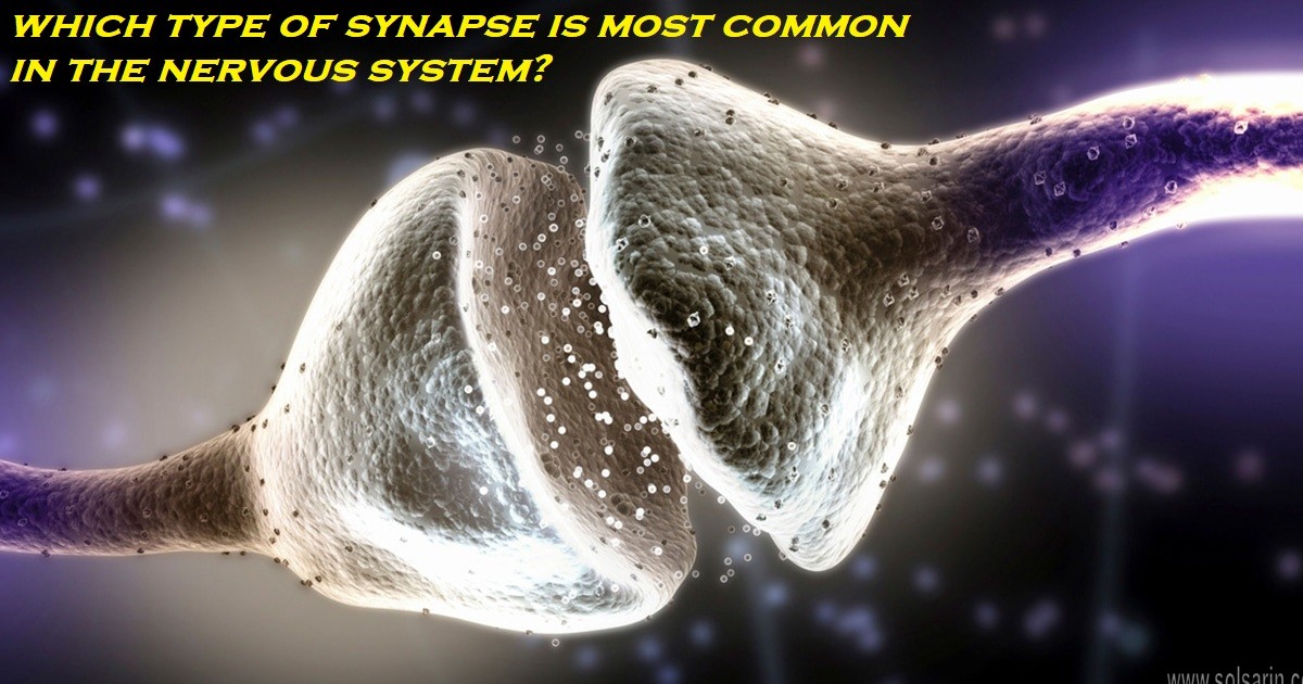
Synapse
In the nervous system, a synapse is a structure that permits a neuron (or nerve cell) to pass an electrical or chemical signal to another neuron or to the target effector cell.
Synapses are essential to the transmission of nervous impulses from one neuron to another. Neurons are specialized to pass signals to individual target cells, and synapses are the means by which they do so. At a synapse, the plasma membrane of the signal-passing neuron (the presynaptic neuron) comes into close apposition with the membrane of the target (postsynaptic) cell.
Both the presynaptic and postsynaptic sites contain extensive arrays of molecular machinery that link the two membranes together and carry out the signaling process. In many synapses, the presynaptic part is located on an axon and the postsynaptic part is located on a dendrite or soma. Astrocytes also exchange information with the synaptic neurons, responding to synaptic activity and, in turn, regulating neurotransmission.[3]
SAMs
Synapses (at least chemical synapses) are stabilized in position by synaptic adhesion molecules (SAMs) projecting from both the pre- and post-synaptic neuron and sticking together where they overlap; SAMs may also assist in the generation and functioning of synapses.[4]
Some authors generalize the concept of the synapse to include the communication from a neuron to any other cell type,[5] such as to a motor cell, although such non-neuronal contacts may be referred to as junctions (a historically older term). A landmark study by Sanford Palay demonstrated the existence of synapses.[6]
Physiology of the Neuron
Krishnagopal Dharani, in The Biology of Thought, 2015
Types of Synapses
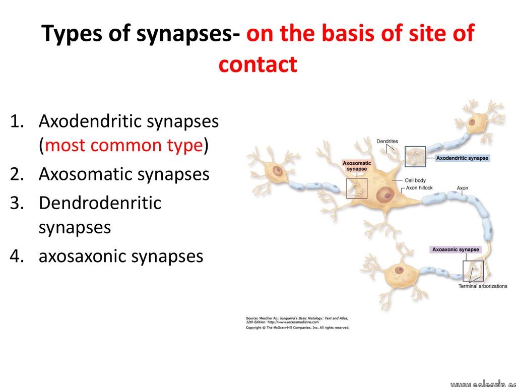
- ●this typically forms when the membrane of the terminal bulb of an axon comes into contact with that of a dendrite (Fig. 2.8). The axodendritic synapses are either excitatory or inhibitory. Axodendritic synapses are the commonest type of synapse in the CNS and are described fully in Chapter 6, p. 110.
- ●here the axon synapses directly with the soma – these may be excitatory or inhibitory.
- ● It connects with axon of another neuron – these are usually inhibitory, as they make the receiving neuron less excitable.
- ●synapse between dendrite and dendrite (Ch. 8, p. 149).
- ● a dendrite communicates with axon (rare).
- ● Synapse between soma and another soma.
- ●an axon may finally end on muscle fibers (commonest synapse in the peripheral nervous system).
CNS
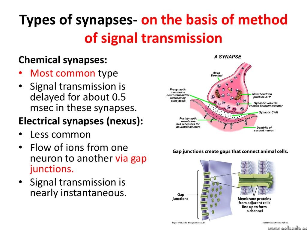
Synapse in the following description means an axodendritic synapse, because they are the commonest synapse in the CNS. In a synapse, the terminal bulb’s membrane forms the presynaptic membrane (Fig. 2.7) and the dendritic membrane of the receiving neuron forms the postsynaptic membrane, and the potential gap between the two is the synaptic cleft (usually about 20–30 nm). The presynaptic membrane (i.e. axon’s membrane) secretes NTs into the synaptic cleft, and the NT acts on the postsynaptic membrane to show the desired effect. The following section describes more about the electrical activity at the synaptic junctions.
The most important function of a chemical synapse is its ability to show synaptic plasticity, and this is the fundamental property of neurons that confers the human brain its capacity for memory and learning, and intelligence – which in turn forms the basis of all higher intellectual functions. This special feature is described in Chapter 3, pp. 67–71.
Type
Silent Synapses
Silent synapses are synapses, which only stably express NMDARs at the postsynaptic site. As discussed above, AMPARs usually accompany NMDARs at the postsynaptic site because NMDARs require an elevated level of depolarization before they are able to function. But in this case, because AMPARs are not stably expressed, when glutamate is released into the synaptic space and on to the NMDARs these specific synapses are nonfunctional – hence the name “silent synapses.”
Silent synapses have shown to induced de novo within the NAc following exposure to cocaine. Evidence shows that silent synapses may be a precursor to a newly generated synapse, existing at a stage during synapse development before AMPARs stably inserted into the membrane to complement the NMDARs.
This line of thought indicates that entirely new circuits and pathways formed within the mesolimbic dopamine system in response to drug experiences. Additionally, synaptic plasticity appears to occur more readily at silent synapses, as these synapses appear primed for LTP-like changes rather than LTD-like changes.
Synaptic Transmission in a Simple Reflex Circuit
One of the simplest behaviors mediated by the central nervous system is knee-jerk or stretch reflex. In response to a neurologist’s hammer to the patella tendon, there is a reflex extension of the leg. Figure 6.1 illustrates the neurocircuitry that controls that reflex response. The stretch to the patella tendon stretches the extensor muscle. More specifically, it stretches a group of specific receptors known as muscle spindle receptors or simply stretch receptors.
The stretch elicits action potentials in the stretch receptors which then propagate over type 1A afferent fibers, the somata of which are located in the dorsal root ganglion. Processes of these sensory neurons then enter the spinal cord and make synaptic connections with two types of cells. First, a synaptic connection formed with the extensor motor neuron located in the ventral horn of the spinal cord.
As the result of synaptic activation of this motor neuron, action potentials elicited in the motor neuron and propagate out the ventral roots, ultimately invading the terminal regions of the motor axon (i.e., the neuromuscular junction), causing release of acetylcholine, depolarization of the muscle cell, formation of an action potential in the muscle cell, and a subsequent contraction of the muscle.
Reflex
The sensory neurons also make synaptic connections with another type of neuron in the spinal cord called an interneuron. Interneurons so named because they interposed between one type of neuron and another. The particular interneuron shown is an inhibitory interneuron. As a result of its activation through the process of synaptic transmission, action potentials elicited in the interneuron.
An action potential in the inhibitory neuron leads to the release of a chemical transmitter substance that inhibits the flexor motor neuron, thereby preventing an improper movement from occurring. This particular reflex known as the monosynaptic stretch reflex because this reflex mediated by a single excitatory synaptic relay in the central nervous system.
In the central nervous system, a synapse is a small gap at the end of a neuron that allows a signal to pass from one neuron to the next. Synapses found where nerve cells connect with other nerve cells.
1897
Synapses are key to the brain’s function, especially when it comes to memory.1
The term synapse first introduced in 1897 by physiologist Michael Foster in his “Textbook of Physiology” and derived from the Greek synapsis, meaning “conjunction.”
What Synapses Do
When a nerve signal reaches the end of the neuron, it cannot simply continue to the next cell. Instead, it must trigger the release of neurotransmitters which can then carry the impulse across the synapse to the next neuron.
Once a nerve impulse has triggered the release of neurotransmitters. These chemical messengers cross the tiny synaptic gap. And taken up by receptors on the surface of the next cell.
These receptors act much like a lock, while the neurotransmitters function much like keys. Neurotransmitters may excite or inhibit the neuron they bind to.
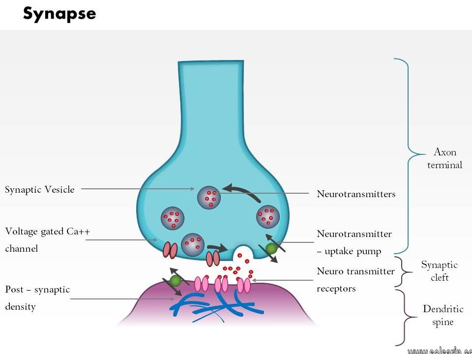
Structure of a chemical synapse. (CC BY).
- The process of synaptic transmission at a chemical synapse between two neurons follows these steps:
- An action potential, propagating along the axon of a presynaptic neuron, arrives at the axon terminal.
- The depolarization of the axolemma at the axon terminal opens Ca2+ channels and Ca2+ diffuses into the axon terminal.
- Ca2+ bind with calmodulin, the ubiquitous intracellular calcium receptor, causing the synaptic vesicles to migrate to. And fuse with the presynaptic membrane.
- The neurotransmitter released into the synaptic cleft by the process of exocytosis.
- The neurotransmitter diffuses across the synaptic cleft and binds with receptors on the postsynaptic membrane.
- Binding of the neurotransmitters to the postsynaptic receptors causes a response in the postsynaptic cell.
Neurotransmitter Effects
The response of the postsynaptic cell to a neurotransmitter depends on the specific receptors. That are present on its cell membrane. Most neurotransmitters can bind to more than one receptor found in the body. And the cell’s response dependent on which receptor bound. Different receptors produce different cellular responses because they activate processes in the cell.
Thank you for staying with this post “which type of synapse is most common in the nervous system?” until the end.